
Sudden swelling in one leg can come from a pulled muscle, infection, a ruptured knee cyst – or a deep vein thrombosis. The safest way to tell them apart is not pain intensity. It is a structured assessment of your risk followed, when needed, by the right blood test or ultrasound.

Patients often tell me, “My legs have always been a little swollen, but lately one is noticeably more swollen than the other.” Some also ask, “Could something be blocked?” I start with a simple question: “When did you first notice the change?” If the difference is not obvious when I examine their legs, I ask about what they notice at home: “Does the swelling go down after you sleep with your legs elevated?” These questions help me understand when the swelling began and how it changes over time.
The uncomfortable question is obvious: Could this be a blood clot?
Sometimes it is. Often it is not. A strained calf muscle, cellulitis, a ruptured Baker’s cyst and several other conditions can look remarkably similar.
What matters is that you cannot reliably separate them by how painful the leg feels. Deep vein thrombosis, or DVT, may cause swelling, tenderness, warmth or discoloration – but some people have few or no symptoms. That is why a new unexplained difference between the legs deserves clinical assessment rather than a home diagnosis. The CDC notes that about half of people with DVT may have no symptoms at all. (1)
When should I go to the emergency room?
One swollen leg by itself usually calls for prompt evaluation, not panic.
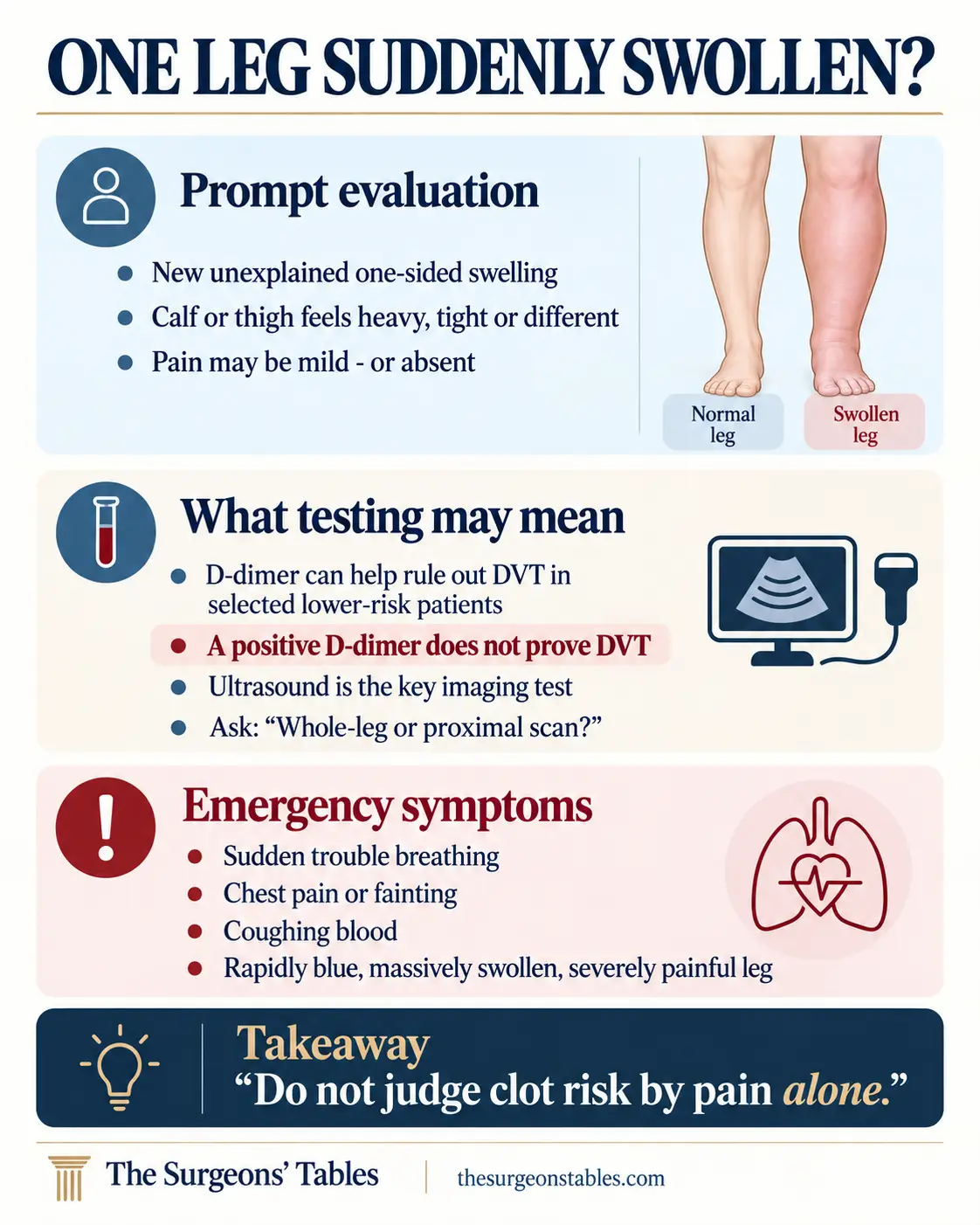
The urgency changes if symptoms suggest that a clot may already have reached the lungs. Seek emergency help for sudden unexplained shortness of breath, chest pain that becomes worse with a deep breath or cough, coughing blood, marked lightheadedness or fainting. A pulmonary embolism can occur even when obvious leg symptoms were never noticed. (1)
There is also a much rarer limb emergency. If the entire leg becomes rapidly and massively swollen, intensely painful and blue or purple – especially if the foot becomes cold, numb, weak or poorly perfused – clinicians worry about phlegmasia, an extreme form of venous obstruction. This needs immediate emergency and vascular-specialist assessment. (2)
Does one swollen leg automatically mean DVT?
No.
One-sided swelling tells a clinician to think about a local problem, but it does not identify which local problem.
| Possible cause | Clues that may fit | What usually separates it |
|---|---|---|
| Deep vein thrombosis | New unilateral swelling, heaviness or tenderness; risk may increase after immobility, surgery, hospitalization, cancer, pregnancy or estrogen exposure. | Clinical probability assessment plus D-dimer and/or compression ultrasound. |
| Calf muscle tear | Sudden focal pain during running, jumping or push-off; bruising and localized tenderness may follow. | History, examination and musculoskeletal imaging when needed. |
| Ruptured Baker’s cyst | Sudden calf swelling or pain, often with an underlying knee problem. | Ultrasound or other imaging can show fluid while confirming that the deep veins remain compressible. |
| Cellulitis | Increasing skin redness, warmth and tenderness, sometimes fever or a visible skin entry point. | Skin-centered examination and clinical evidence of infection rather than deep venous obstruction. |
| Superficial thrombophlebitis | Tender, inflamed superficial vein that may feel like a cord. | Ultrasound can define superficial clot and assess the deep veins when indicated. |
| Pelvic or iliac venous obstruction | Marked unilateral swelling, sometimes involving much of the leg; extensive left-sided DVT can raise suspicion for iliac-vein compression. | Pelvic venous imaging when the clot distribution or clinical pattern points above the leg. |
Why doesn’t the doctor diagnose DVT from my symptoms?
Because the symptoms are not specific enough.
Clinicians first estimate pretest probability – how likely DVT appears before the definitive test is performed. A validated tool such as the Wells rule can contribute to that estimate.
The Wells rule includes findings such as leg swelling, calf-size difference, localized venous tenderness, recent immobilization or surgery, active cancer, previous DVT and whether another diagnosis appears at least as likely.
It is useful as a clinical decision tool. It is not a home diagnostic quiz.
That distinction matters because the next test depends partly on the probability estimated before testing. (3) (4)
What does a D-dimer actually tell me?
A D-dimer test looks for fragments produced when the body forms and breaks down fibrin.
Its great strength is sensitivity, not specificity.
In a suitably low-risk patient, a negative high-sensitivity D-dimer can make DVT unlikely enough that ultrasound may not be necessary. ASH endorses this type of strategy for low-pretest-probability patients. Large diagnostic studies have also shown very low rates of subsequent thromboembolic events after DVT is excluded using clinical probability plus an appropriate negative D-dimer. (3) (5) (6)
But the reverse is not true.
D-dimer can rise with many conditions, including inflammation, recent surgery, trauma and pregnancy, and tends to become less specific with age.
That is why ASH specifically recommends against diagnosing DVT from a positive D-dimer alone. (3)
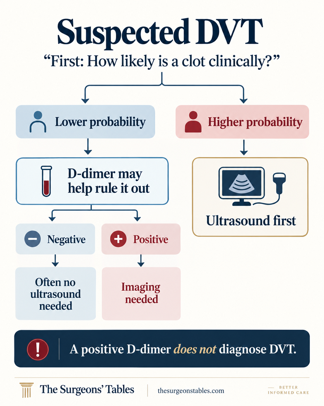
A negative D-dimer can be powerful in the right patient. A positive D-dimer does not prove there is a blood clot.
Why did someone else get an ultrasound immediately?
Because the pretest probability may have been different.
When DVT is clinically more likely, ASH recommends beginning with either proximal or whole-leg ultrasound rather than trying to settle the diagnosis with a positive D-dimer. (3)
Ultrasound looks for a vein that fails to compress normally and for other evidence of thrombosis.
But there is a detail many patients never hear: Not every “negative leg ultrasound” represents the same examination.
Why might I need another ultrasound in a week?
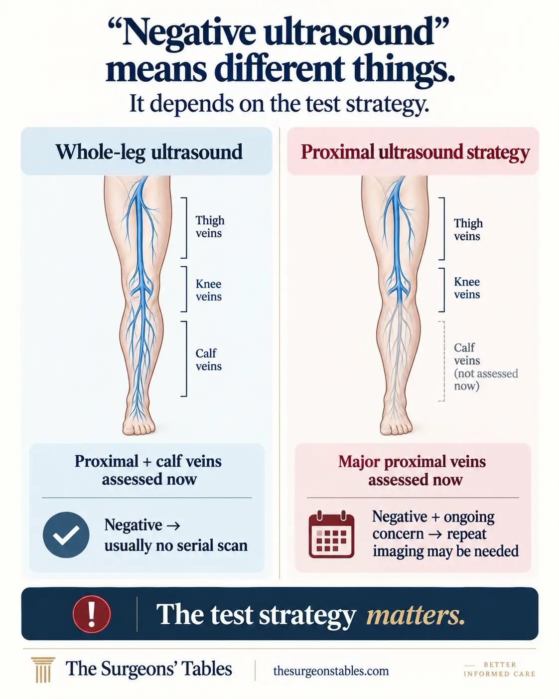
A whole-leg ultrasound evaluates both proximal veins and the calf veins during the initial examination.
A proximal ultrasound concentrates on the veins where a clot carries greater clinical consequences. It can intentionally leave distal calf-vein thrombosis outside the definitive first examination.
That is not necessarily an incomplete test. It can be part of a planned diagnostic strategy.
ASH accepts either whole-leg ultrasound or a proximal-ultrasound strategy in appropriate patients. When an initial proximal study is negative but clinical probability remains meaningful and no better diagnosis explains the symptoms, serial proximal ultrasound may be required. NICE specifies repeat proximal imaging 6 to 8 days later in its relevant pathway. (3) (4)
So if you are told, “The scan is negative, but come back next week,” that does not automatically mean someone mistrusts the first sonographer.
The second scan may be an intentional part of the test.
If a clot is found, why isn’t every DVT treated the same way?
Location matters.
A proximal DVT involves the popliteal, femoral, iliac or other proximal deep veins. When acute VTE is treated with anticoagulation, CHEST recommends a minimum 3-month treatment phase, followed by reassessment of whether therapy should stop or continue longer. (7)
An isolated distal DVT is confined to the calf veins.
For selected patients with an acute isolated distal DVT who do not have severe symptoms or risk factors for extension, CHEST suggests serial ultrasound surveillance over two weeks instead of immediate anticoagulation. If the thrombus extends, particularly into the proximal veins, treatment changes. (7)
This explains a situation that can otherwise look inconsistent: one patient with DVT leaves with an anticoagulant prescription, while another is scheduled for another ultrasound.
They may have two anatomically different diseases carrying different balances of clot progression and bleeding risk.
What if the swelling is mostly on the left and the clot is very extensive?
That can raise a second question: Is something higher in the pelvis obstructing venous drainage?
One possible cause is May-Thurner syndrome, in which the left common iliac vein is compressed in the pelvis. A standard leg ultrasound can diagnose a resulting DVT but may not fully characterize the pelvic anatomy.
When the clinical pattern suggests iliac obstruction, clinicians may use pelvic CT or MR imaging, venography, and in selected interventional settings intravascular ultrasound. IVUS can identify iliofemoral obstructive lesions that venography alone may underestimate. (8) (9)
This is not a test everyone with a swollen left calf needs. It becomes relevant when the clot distribution, severity or patient history points higher than the calf.
What should I do with a newly swollen leg?
Seek prompt medical assessment if one leg develops unexplained new swelling, particularly if the difference is substantial, persists, or is accompanied by clot risk factors.
Do not rely on pain severity, redness, a massage test, exercise history or an internet Wells calculator to clear yourself.
When you are evaluated, tell the clinician about recent surgery or hospitalization, prolonged immobility or travel, previous clots, cancer, pregnancy or postpartum status, estrogen-containing medications, significant injuries and any family history that may be relevant.
And ask what kind of ultrasound was performed.
- Was my clinical probability of DVT considered before testing?
- If I had a D-dimer, was it being used to rule out DVT rather than diagnose it?
- Was my ultrasound a whole-leg or proximal study?
- If the proximal scan was negative, do I need repeat imaging?
- If a clot was found, is it distal or proximal?
Bottom Line
A suddenly swollen leg is not automatically a blood clot, but mild pain does not make DVT safe to ignore.
The useful question is not “Does this hurt enough to be a clot?” It is whether your history and examination put you in a group that can be safely ruled out with a validated D-dimer strategy or whether you need ultrasound.
If you have new unexplained one-sided swelling, seek timely clinical assessment. If you also develop shortness of breath, chest pain, fainting, coughing blood, or a rapidly blue and massively swollen limb, treat that as an emergency.
References
- (1) Centers for Disease Control and Prevention. About Venous Thromboembolism (Blood Clots). Updated March 5, 2025. CDC.
- (2) Kakkos SK, Gohel M, Baekgaard N, et al. European Society for Vascular Surgery 2021 Clinical Practice Guidelines on the Management of Venous Thrombosis. European Journal of Vascular and Endovascular Surgery. 2021;61(1):9-82. PMID: 33334670. PubMed.
- (3) Lim W, Le Gal G, Bates SM, et al. American Society of Hematology 2018 guidelines for management of venous thromboembolism: diagnosis of venous thromboembolism. Blood Advances. 2018;2(22):3226-3256. Original guideline.
- (4) National Institute for Health and Care Excellence. Venous thromboembolic diseases: diagnosis, management and thrombophilia testing. NICE guideline NG158. Current recommendations.
- (5) Wells PS, Anderson DR, Rodger M, et al. Evaluation of D-dimer in the diagnosis of suspected deep-vein thrombosis. New England Journal of Medicine. 2003;349(13):1227-1235. PMID: 14507948. DOI: 10.1056/NEJMoa023153. PubMed.
- (6) Geersing GJ, Zuithoff NPA, Kearon C, et al. Exclusion of deep vein thrombosis using the Wells rule in clinically important subgroups: individual patient data meta-analysis. BMJ. 2014;348:g1340. PMID: 24615063. DOI: 10.1136/bmj.g1340. Original article.
- (7) Stevens SM, Woller SC, Kreuziger LB, et al. Antithrombotic Therapy for VTE Disease: Second Update of the CHEST Guideline and Expert Panel Report. Chest. 2021;160(6):e545-e608. CHEST guideline.
- (8) Society for Vascular Surgery. May-Thurner Syndrome. Society for Vascular Surgery.
- (9) Gagne PJ, Tahara RW, Fastabend CP, et al. Venography versus intravascular ultrasound for diagnosing and treating iliofemoral vein obstruction. PubMed.
- (10) Kearon C, de Wit K, Parpia S, et al. Diagnosis of deep vein thrombosis with D-dimer adjusted to clinical probability: prospective diagnostic management study. BMJ. 2022;376:e067378. DOI: 10.1136/bmj-2021-067378. Original article.