
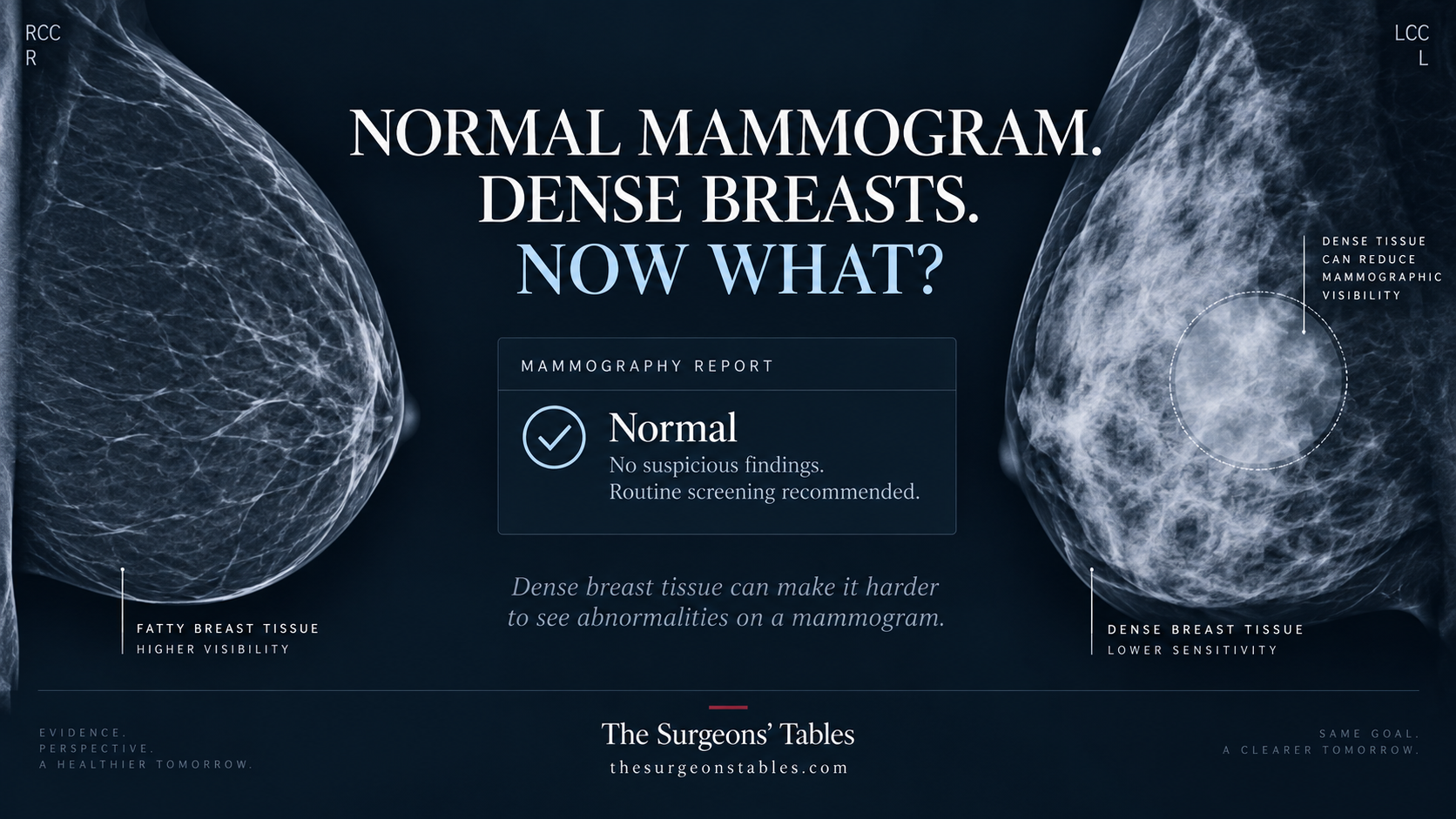
Your mammogram was normal. So why does the dense-breast letter say cancer could still be missed? The answer is not automatically another scan.
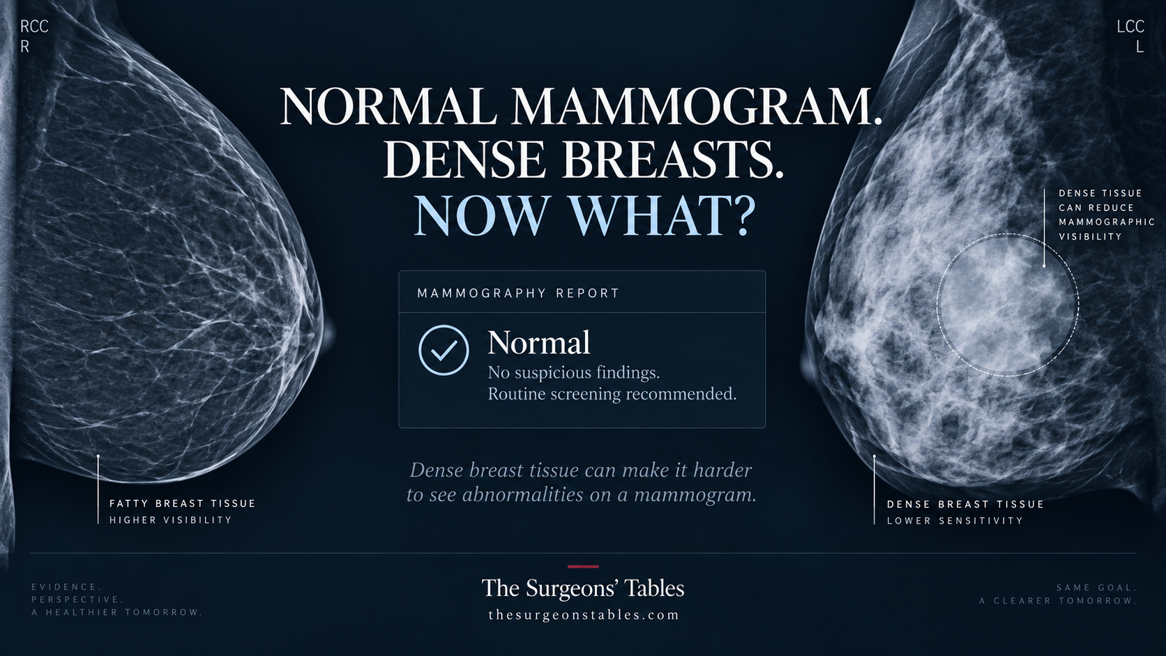
Your mammogram was normal.
Then the letter told you that your breasts are dense – and that dense tissue can make cancer harder to see.
So which message are you supposed to believe?
Both.
A negative screening mammogram means the radiologist did not see evidence of cancer on that examination. Dense breast tissue means mammography may be less sensitive in your breasts.
Those statements are not contradictory.
And neither one automatically means you need an ultrasound or MRI.
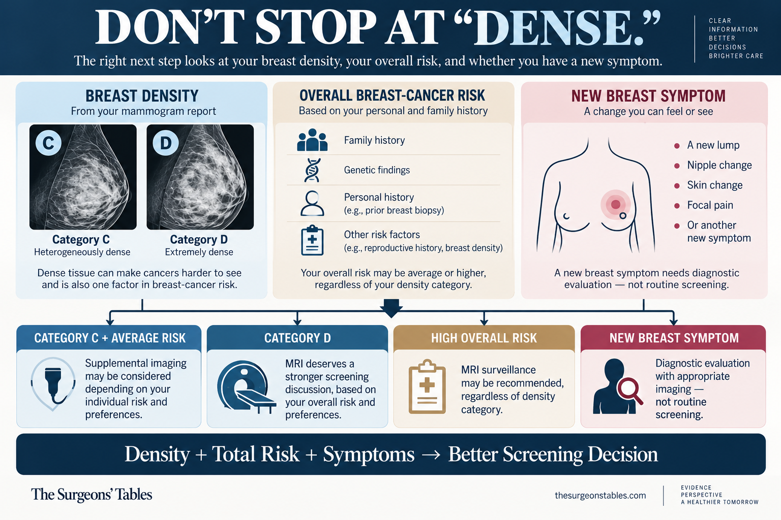
The real decision depends mainly on three things:
How dense are your breasts? What is your overall breast-cancer risk? And what would another test add – including the chance that it starts a false-positive workup?
Why have a mammogram if I might need an ultrasound too?
When patients learn they have dense breasts, a common frustration I hear is, “If I’m going to need an ultrasound or another test anyway, why do I have to go through a painful mammogram? Can’t I just skip it?” Some also ask, “If dense tissue makes things hard to see, won’t the other tests have the same problem?”
I explain that mammography and ultrasound are not simply cheaper and more expensive versions of the same test. They look at breast tissue in different ways. I compare them to tools designed for different viewing conditions: the useful tool depends on what we need to see.
Mammography can reveal tiny calcium deposits that ultrasound may not show. Ultrasound can help evaluate findings that are difficult to see on a mammogram. The tests can complement each other, but dense breasts do not automatically mean everyone needs both. (7); (8).
My report says Category C or D. Does that actually change anything?
Yes.
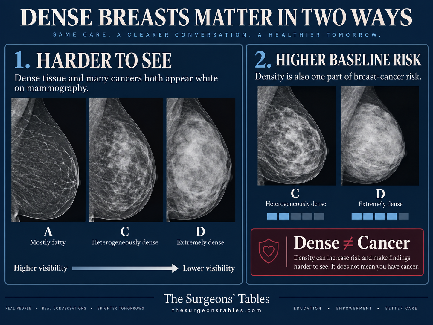
Breast density describes how much fibrous and glandular tissue appears on a mammogram compared with fat. Fat appears relatively dark. Dense tissue appears white.
The problem is that many breast cancers also appear white. A small cancer can therefore be harder to distinguish when the surrounding breast tissue is very dense.
Radiologists divide breast density into four BI-RADS categories. The two considered dense are C and D.
| Category | What it means | Why it matters |
|---|---|---|
| C: Heterogeneously dense | Dense areas may obscure small masses. | Supplemental imaging may be considered, particularly if other risk factors are present. |
| D: Extremely dense | Mammographic sensitivity is reduced more substantially. | The case for supplemental MRI becomes stronger. |
Category C and Category D should not be treated as the same situation.
For an otherwise average-risk woman with Category C breasts, the ACR considers ultrasound, contrast-enhanced MRI and abbreviated MRI possible supplemental options rather than automatic requirements. (9)
For an average-risk woman with Category D breasts, the ACR rates contrast-enhanced MRI and abbreviated MRI as usually appropriate. (9)
That does not make MRI mandatory. It means extremely dense tissue changes the balance enough that MRI deserves a more serious discussion.
Dense breasts themselves are not cancer, a lump or a precancerous condition. They are common anatomy.
But density matters for two separate reasons: it can hide cancer on mammography, and it is also associated with increased breast-cancer risk.
Does the federal dense-breast letter mean the government thinks I need another scan?
No.
Since September 10, 2024, mammography facilities across the United States have been required to tell patients whether their breasts are dense. (10)
The FDA notification explains that dense tissue can make cancer harder to find and that, in some people, additional imaging may help.
Those words matter.
The rule requires that patients be informed. It does not require every woman with dense breasts to undergo an ultrasound or MRI.
The USPSTF concluded in 2024 that there is still insufficient evidence to determine the overall balance of benefits and harms of supplemental ultrasound or MRI in women with dense breasts and an otherwise negative mammogram. (11)
That “I statement” is not a recommendation against additional screening. It means the Task Force does not believe the evidence is strong enough to recommend routinely for or against it for everyone.
ACOG also does not recommend routine extra screening when dense breasts are the only identified risk factor. (12)
So receiving the letter is a reason to look more closely at your risk – not an automatic instruction to schedule another scan.
What may matter even more than breast density?
Your overall breast-cancer risk.
Consider two women whose mammograms both say Category C.
One has no strong family history, no known high-risk genetic variant and no previous high-risk breast biopsy.
The other has a strong family history and a calculated lifetime breast-cancer risk of 25%.
Their density label is identical. Their screening plans may not be.
The ACR recommends MRI surveillance for women with a calculated lifetime risk of 20% or more, as well as several other higher-risk groups. (1)
That creates an important clinical reversal:
A woman with Category C breasts and substantial additional risk may have a stronger indication for MRI than another woman whose only known risk factor is dense tissue.
Family history, genetic variants, personal history of breast cancer, prior chest radiation and certain previous breast-biopsy findings can all affect risk.
Risk calculators such as Tyrer-Cuzick are often used, but no risk score should be interpreted in isolation. The model has to fit the patient and use accurate family and medical history.
The practical sequence is: First assess risk. Then decide whether supplemental imaging adds enough value.
If MRI finds more cancers, why would I be offered ultrasound?
Because sensitivity is not the only thing that determines which test makes sense.
Ultrasound has several advantages. It does not use ionizing radiation, it does not require intravenous contrast, and it is generally easier to access.
It can also find cancers that mammography misses.
The trade-off is that it frequently finds abnormalities that are not cancer.
In a large U.S. Breast Cancer Surveillance Consortium study, supplemental ultrasound detected about 2 cancers per 1,000 examinations overall. Its cancer-detection rate was higher in women who were already at higher underlying risk. (5)
That last detail matters.
The value of the same test changes with the risk of the person being tested.
MRI is more sensitive.
The randomized DENSE trial studied women ages 50 to 75 with extremely dense breasts and a negative mammogram. Women invited to supplemental MRI had about 2.5 interval cancers per 1,000 screenings, compared with 5.0 per 1,000 in the mammography-only group. (2)
An interval cancer is one diagnosed after a negative screening examination but before the next scheduled screen. Reducing interval cancers is clinically meaningful.
But MRI also produced false positives.
During the first DENSE MRI round, there were roughly 80 false-positive examinations per 1,000 screenings. In the second round, once previous MRI images were available for comparison, that fell to about 26 per 1,000. (2), (3)
That is a useful point many patients never hear: The false-alarm burden of breast MRI may be greatest the first time it is performed.
The newer BRAID randomized trial also found a substantial detection difference. In its first supplemental screening round, abbreviated MRI detected 17.4 cancers per 1,000 examinations, compared with 4.2 per 1,000 with automated whole-breast ultrasound. (4)
But that does not settle the entire question.
Finding more cancers is not automatically the same as proving that a screening strategy reduces breast-cancer deaths. BRAID also did not establish how much overdiagnosis will ultimately result.
So MRI’s higher detection rate is important – but it is not a reason to tell every woman with dense breasts that MRI is mandatory.
| Feature | Ultrasound | MRI / abbreviated MRI |
|---|---|---|
| Finds cancers mammography misses | Yes | Yes, generally more |
| IV contrast | No | Usually yes |
| Ionizing radiation | No | No |
| False-positive findings | Common | Also occur, particularly on the first MRI |
| Availability | Usually easier | More variable |
| Cost | Generally lower | Generally higher |
| Strongest role | Selected dense-breast patients; alternative when MRI is unavailable or unsuitable | High-risk patients and women for whom extreme density substantially limits mammography |
| Proven mortality benefit from supplemental screening alone | Not established | Not established |
So if ultrasound is recommended, the useful issue is not whether ultrasound is a “good” or “bad” test.
It is whether ultrasound is the right supplemental test for this particular risk profile and clinical situation.
MRI may not be locally available. Insurance coverage may differ. Some patients cannot tolerate the examination. Others may have contrast or device considerations.
But the easiest test to obtain and the most informative test are not always the same thing.
What does a “false alarm” actually mean for me?
This deserves more explanation than a percentage.
An additional scan does not produce only two possible answers: “cancer” or “nothing.”
It may find something uncertain.
That can start a sequence such as:
supplemental scan -> uncertain finding -> targeted imaging or additional views -> short-term follow-up or biopsy -> often a benign result
A benign biopsy does not necessarily mean the original test was a mistake. False positives are one consequence of trying to increase sensitivity and find cancers that mammography may miss.
But the burden is real. There can be additional appointments, anxiety, procedure discomfort and out-of-pocket costs.
That trade-off will not feel the same to every patient.
A woman at high risk may reasonably accept a greater chance of benign workup in exchange for higher sensitivity.
For an otherwise average-risk woman with Category C breasts, that same trade-off may be much more preference-sensitive.
This is why “MRI finds more cancers” cannot be the only sentence used to make the decision.
A dense-breast notification tells you that mammography may have a blind spot. It does not tell you, by itself, whether another scan is worth chasing that blind spot.
Why can two good doctors give different recommendations?
Because professional groups are using different thresholds for acting on incomplete evidence.
| Organization | General approach | What it means |
|---|---|---|
| USPSTF | Evidence is insufficient to routinely recommend for or against supplemental US or MRI after a negative mammogram in dense breasts. | No universal additional-test recommendation. |
| ACOG | Does not recommend routine extra screening when dense breasts are the only identified risk factor. | Density alone may not justify another test. |
| ACR | Uses density and overall risk; MRI becomes more strongly favored in extremely dense and higher-risk breasts. | Category C and D are not treated identically. |
| EUSOBI | Recommends supplemental MRI for extremely dense breasts; ultrasound may be used when MRI is unavailable. | More proactive approach to Category D. |
The disagreement is not simply about whether MRI detects additional cancer. It clearly does.
The harder question is whether routinely adding imaging for every dense-breast patient improves long-term outcomes enough to justify more recalls, biopsies, contrast exposure, cost and possible overdiagnosis.
That remains uncertain.
What about gadolinium if I have kidney disease?
Standard screening breast MRI usually uses intravenous gadolinium contrast.
A previous severe contrast reaction, certain implanted devices, severe claustrophobia, acute kidney injury or advanced kidney disease can affect how MRI is planned.
But the old shorthand that an eGFR below 30 automatically means “no gadolinium” is too simplistic.
With modern group II gadolinium agents, the risk of nephrogenic systemic fibrosis is considered very low even in advanced kidney disease. (13)
That does not mean patients should make the contrast decision themselves. It means severe kidney disease should prompt an individualized discussion with the radiology team rather than an automatic assumption that breast MRI is impossible.
What if my mammogram was normal but I can feel a lump?
Then this is no longer mainly a dense-breast screening question.
It becomes a diagnostic question.
A new palpable lump, spontaneous suspicious nipple discharge, new nipple or skin retraction, or another persistent focal breast change deserves diagnostic evaluation even if the last screening mammogram was negative.
The exact first test depends partly on age and the finding.
For a woman 40 or older with a new palpable mass, diagnostic mammography or tomosynthesis is generally appropriate, often followed by targeted ultrasound.
For a woman younger than 30 with a palpable mass, ultrasound is usually the initial imaging examination. (6)
The principle is more important than memorizing the age cutoffs: A normal screening mammogram does not cancel a new physical finding.
So what should I actually do after getting the letter?
Do not start by automatically booking an ultrasound.
1. Read the density category.
Category C and Category D are not the same clinical situation.
2. Put density into your total risk profile.
Family history, genetics, personal history and previous biopsy findings may matter more than the density letter itself. If calculated lifetime risk is 20% or higher, MRI may be recommended regardless of density.
3. Match the supplemental test to the reason for testing.
Category D and higher-risk patients have a stronger case for MRI. Ultrasound remains useful when MRI is unavailable, unsuitable or chosen for other individual reasons.
4. Know what happens after an uncertain result.
More sensitive imaging can mean more recalls, additional scans and sometimes benign biopsies. That is part of the decision, not an afterthought.
5. Check practical barriers before scheduling elective supplemental screening.
Insurance coverage, local MRI availability and follow-up costs can matter.
And if there is a new breast symptom, skip this screening decision tree altogether and pursue diagnostic evaluation.
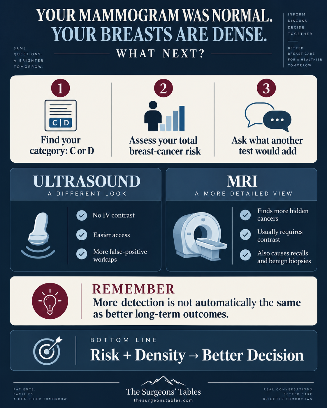
Bottom Line
A dense-breast notification tells you that mammography may have a blind spot.
It does not tell you, by itself, whether another scan is worth chasing that blind spot.
For an otherwise average-risk woman with Category C breasts, supplemental imaging is often a preference-sensitive decision.
For Category D, the MRI discussion becomes more compelling.
And when overall breast-cancer risk is high, MRI may be recommended regardless of density.
The decision becomes much clearer when three things are known: your density category, your total breast-cancer risk, and what you are willing to do with an uncertain result.
References
- (1) Monticciolo DL, et al. Breast Cancer Screening for Women at Higher-Than-Average Risk: Updated Recommendations From the ACR. J Am Coll Radiol. 2023;20(9):902-914. DOI 10.1016/j.jacr.2023.04.002. PMID 37150275. PubMed.
- (2) Bakker MF, et al. Supplemental MRI Screening for Women with Extremely Dense Breast Tissue. N Engl J Med. 2019;381(22):2091-2102. DOI 10.1056/NEJMoa1903986. PMID 31774954. PubMed.
- (3) Veenhuizen SGA, et al. Supplemental Breast MRI for Women with Extremely Dense Breasts: Results of the Second Screening Round of the DENSE Trial. Radiology. 2021;299(2):278-286. DOI 10.1148/radiol.2021203633. PMID 33724062. PubMed.
- (4) Gilbert FJ, et al. Comparison of supplemental breast cancer imaging techniques – interim results from the BRAID randomised controlled trial. Lancet. 2025;405(10493):1935-1944. DOI 10.1016/S0140-6736(25)00582-3. PMID 40412427. PubMed.
- (5) Sprague BL, et al. Performance of Supplemental US Screening in Women with Dense Breasts and Varying Breast Cancer Risk: Results from the Breast Cancer Surveillance Consortium. Radiology. 2024;312(2):e232380. DOI 10.1148/radiol.232380. PMID 39105648. PubMed.
- (6) American College of Radiology. ACR Appropriateness Criteria: Palpable Breast Masses. ACR.
- (7) ACR/RSNA: Breast Ultrasound
- (8) NCI: Dense Breasts
- (9) Paulis LV, et al. ACR Appropriateness Criteria Supplemental Breast Cancer Screening Based on Breast Density: 2024 Update. J Am Coll Radiol. 2025;22(5S):S405-S423. DOI 10.1016/j.jacr.2025.02.023. PMID 40409891. PubMed.
- (10) U.S. Food and Drug Administration. Mammography Quality Standards Act Final Rule and breast density notification requirements. FDA.
- (11) U.S. Preventive Services Task Force. Screening for Breast Cancer. 2024. USPSTF.
- (12) American College of Obstetricians and Gynecologists. Dense Breasts. Last reviewed May 2026. ACOG.
- (13) Weinreb JC, et al. Use of Intravenous Gadolinium-Based Contrast Media in Patients With Kidney Disease: Consensus Statements from the American College of Radiology and the National Kidney Foundation. Radiology. 2021;298(1):28-35. PMID 33170103. PubMed.
- (14) Marcon M, Fuchsjager MH, Clauser P, Mann RM. ESR Essentials: screening for breast cancer – general recommendations by EUSOBI. Eur Radiol. 2024;34(10):6348-6357. DOI 10.1007/s00330-024-10740-5. PMID 38656711. PubMed.